🌍
Global International Hub
🗨️
👨‍⚕️
Aspen Assistant Online
Hi! Welcome to Aspen Dental Care. Looking for a brighter smile? How can I help you today?
⭐ Rated 5.0 on Google — 200+ verified reviews 🦷 20+ Years of Trusted Dental Care in RT Nagar 💬 Raveena just booked a Dental Implant consultation ✅ Free first consultation — No commitment required 💳 EMI available · 0% interest on select plans 🏥 20+ dental specialities under one roof 📞 We respond within 1 hour on WhatsApp 🦷 Priya saved ₹8,000 with our EMI plan this week ⭐ Rated 5.0 on Google — 200+ verified reviews 🦷 20+ Years of Trusted Dental Care in RT Nagar 💬 Raveena just booked a Dental Implant consultation ✅ Free first consultation — No commitment required 💳 EMI available · 0% interest on select plans 🏥 20+ dental specialities under one roof 📞 We respond within 1 hour on WhatsApp 🦷 Priya saved ₹8,000 with our EMI plan this week
📅 Book Appointment
Microscopic Dentistry

See More.
Treat Better.
Heal Faster.

At Aspen Dental Care, every single treatment — from a root canal to a veneer preparation — is performed under an advanced dental operating microscope. Magnification from 5× to 25× reveals what the naked eye simply cannot see.

25× Magnification
100% Microscope-Guided
🦷
Viewing at 16× Magnification
🔬
Dental Microscope
5× – 25× zoom
💡
Coaxial Illumination
Shadow-free light
📹
Video Documentation
4K live capture
🎯
Precision Treatment
Sub-mm accuracy
Visibility improved by 2500%
25×Max Magnification
100%Microscope-Guided
20+Yrs Clinical Experience
4KLive Video Documentation

The Technology

What is
Microscopic Dentistry?

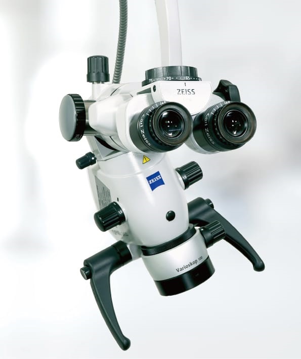
Microscopic dentistry uses a high-powered dental operating microscope (DOM) mounted on an articulating arm to illuminate and magnify the treatment field up to 25×. What looks like a uniform surface to the naked eye reveals intricate anatomy — accessory canals, micro-fractures, hidden decay margins, and tissue planes — under magnification.

At Aspen Dental Care, this is not an add-on for complex cases. Every treatment, for every patient, is performed under the microscope. It is our standard of care — because precision shouldn't be optional.

Operative Dentistry
Root Canal Therapy
Cosmetic Procedures
Periodontal Surgery
Implant Placement
Dental Treatment

Your Visit

How a Microscope-Guided
Appointment Works

From first consultation to final follow-up, the microscope is central to every step.

🩺
Microscopic Examination
We examine your teeth, gums, and restorations under magnification — detecting issues invisible to the naked eye before they become bigger problems.
🎯
Precise Diagnosis
Micro-fractures, secondary decay, hidden canals, and tissue anatomy are clearly visualised and recorded in 4K video — shared with you in real time.
🔬
Microscope-Guided Treatment
Every instrument movement — whether a scalpel, file, or bur — is guided by the magnified view, ensuring sub-millimetre accuracy throughout.
Verified Outcome
Before you leave, we use the microscope to verify the final result — seal integrity, margin fit, canal obturation quality, suture placement — nothing is assumed.

Our Services

Precision in,
Every Procedure

We don't reserve the microscope for "difficult" cases. It is used for every procedure — giving every patient the highest possible standard of care.

🦷
Microscopic Root Canal (RCT)
Our Speciality

Root canals are perhaps the single greatest beneficiary of microscopy. At 25× magnification, accessory canals, calcified passages, missed canals, and fins — invisible otherwise — are located, cleaned, and sealed with certainty. The result: a root canal that is thorough, comfortable, and built to last decades.

Detection of all root canal anatomy including accessory & lateral canals
Identification and removal of fractured instruments under vision
Conservative tooth structure removal — only diseased tissue targeted
Verified obturation quality before appointment ends
🪥
Composite Fillings & Bonding
Operative Dentistry

Under the microscope, even the smallest cavity is fully excavated without touching healthy enamel. Bonding layers are applied with perfect adaptation, and the final restoration is sculpted and polished to a seamless finish.

Complete caries removal confirmed visually — no guesswork
Perfect margin sealing reducing secondary decay risk
Aesthetic contouring at micro-level for natural translucency
Porcelain Veneers
Cosmetic Dentistry

Veneer preparation demands precision at every micron. The microscope allows us to prepare the minimal enamel necessary, verify perfect margin placement, and ensure the final porcelain bond is optimal — for veneers that last a lifetime.

Ultra-conservative preparation — preserving maximum tooth structure
Precise chamfer margin for invisible veneer edges
Perfect bonding protocol verified under magnification
👑
Crowns & Bridges
Restorative Dentistry

Crown preparation under the microscope means precisely controlled taper, perfectly placed margins at or just below the gumline, and a prep design that maximises retention while protecting the pulp. Temporary and permanent crowns are fitted under visual verification.

Sub-gingival margins placed with controlled depth
Pulp proximity monitored in real time
Final seating and cement removal confirmed microscopically
🌿
Periodontal & Gum Surgery
Periodontology

Microsurgical periodontal techniques use finer instruments and smaller incisions than traditional surgery, resulting in dramatically less pain, swelling, and healing time. Flap design, root debridement, and suturing are all performed at microscope level.

Minimally invasive flap incisions for faster healing
Complete calculus removal verified at magnification
Micro-suturing for minimal scarring
🔩
Dental Implants
Implantology

Microscope-guided implant surgery ensures the osteotomy site is prepared with optimal angulation, precise depth, and perfect relation to adjacent anatomy. Implant placement, abutment seating, and soft tissue management are all performed under visual control.

Optimal implant positioning with microscope guidance
Abutment-crown margins verified for perfect fit
Soft tissue architecture sculpted for natural emergence
🩹
Atraumatic Extractions
Oral Surgery

Under the microscope, the periodontal ligament space is clearly visible, allowing instruments to be guided precisely between the root and alveolar bone — preserving the socket and surrounding bone for future implant placement.

Socket preservation for future implant readiness
Minimal trauma to surrounding gum and bone
Reduced post-operative discomfort
👶
Paediatric Dentistry
Kids Dentistry

Children's teeth have thinner enamel and closer pulp chambers — making precision even more critical. Microscopy allows us to treat children with minimal intervention, reducing anxiety, chair time, and the risk of accidental pulp exposure.

Ultra-conservative fillings in primary teeth
Pulpotomy and pulpectomy under microscope for better outcomes
Less time in the chair means less anxiety for young patients
💎
Teeth Whitening & Smile Design
Aesthetic Dentistry

Before any whitening or smile makeover, we use the microscope to assess enamel integrity, existing restoration margins, and gum health — ensuring the treatment plan is safe and will deliver predictable results.

Enamel surface assessment before bleaching agents are applied
Gingival tissue protection verified under magnification
Post-treatment surface quality checked microscopically

Why it Matters

Magnification Changes
Everything

The level of visual detail directly determines the quality of treatment possible. See how we compare.

Naked Eye
Gross anatomy visible
Micro-fractures missed
Accessory canals unseen
Sub-marginal decay hidden
Tissue planes indistinct
No documentation
3.5×
Dental Loupes
Improved overall view
Better posture for dentist
Limited depth of field
Accessory canals still missed
No variable magnification
No video documentation
5–25×
Dental Microscope
All anatomy clearly visible
Micro-fractures detected
All canal anatomy found
Hidden decay revealed
Variable magnification
4K video documentation

Patient Benefits

What You Gain with
Microscopic Dentistry

🎯
Unrivalled Precision

Every cut, every fill, every canal is performed at sub-millimetre accuracy. The microscope transforms dentistry from an art of estimation into a science of certainty.

🛡️
More Tooth Conservation

By seeing exactly what needs to be removed, we take only diseased tissue — preserving healthy enamel, dentine, and pulp. Your natural tooth stays stronger, longer.

💊
Less Pain, Faster Healing

Microsurgical techniques use smaller incisions, create less tissue trauma, and allow more controlled tissue handling — translating to significantly less post-operative discomfort.

📹
See What We See

The microscope connects to a 4K monitor so you can see your own treatment in real time. No more mystery — just clear, transparent dentistry where you are fully informed.

♾️
Longer-Lasting Results

Restorations placed with microscope precision have better marginal seal, superior aesthetics, and longer clinical longevity — saving you time, money, and repeat procedures.

🔍
Earlier Problem Detection

Conditions that would be missed at naked-eye examination — micro-cracks, early interproximal lesions, cracked cusp syndrome — are identified and managed before they escalate.

Your Journey

Your Microscope-Guided
Patient Experience

From the moment you walk in to the moment you leave, our microscope-first approach means you receive treatment that is more thorough, more comfortable, and more transparent than you have ever experienced.

🔬
See Your Own Diagnosis
Live microscope feed on the patient monitor shows exactly what we find — no guesswork, no uncertainty.
📋
Video Treatment Records
Your treatment is documented in 4K. You receive a record of your procedure for complete transparency.
🤝
Informed Consent
You see what we see before we treat. Treatment is only begun once you fully understand and agree.
Arrival & Welcome
You are welcomed and your history reviewed. The microscope is positioned and calibrated for your appointment.
First 10 min
Microscopic Examination
We examine all teeth and soft tissue under magnification. Any findings are shown to you on the monitor in real time.
15–20 min
Treatment Discussion
We review what the microscope revealed, walk through your options, and answer every question — before touching a single instrument.
10–15 min
Microscope-Guided Treatment
Treatment proceeds under full microscope vision. Every step is performed with the precision and control only magnification can provide.
As required
Verified Outcome
The completed work is inspected under the microscope before you leave. We confirm everything is perfect — not assumed.
Final 10 min

Questions

Frequently Asked
Questions

Is every treatment really done under the microscope? +
Yes — without exception. At Aspen Dental Care, the dental operating microscope is used for every patient, for every procedure. Whether you are here for a routine filling, a root canal, a veneer, or a periodontal surgery, your treatment will be performed under magnification. This is our standard of care, not an optional upgrade.
Does microscopic dentistry cost more? +
Microscopic dentistry at Aspen Dental Care is priced to be accessible. While the technology investment is significant, we believe patients deserve precision as a standard — not a luxury. We also offer convenient EMI options with 0% interest on select plans, making high-quality care within reach for every family.
Will I feel anything different during a microscope-guided treatment? +
You will experience less discomfort, not more. The microscope allows us to be more precise and less invasive — smaller incisions, targeted tissue removal, and better anaesthesia placement. Most patients report that procedures feel significantly more comfortable than at their previous practice. The main visible difference is the monitor showing you your own treatment in real time.
My previous root canal failed. Can a microscope help re-treat it? +
Absolutely — and this is one of the strongest applications of microscopy. Failed root canals are commonly due to missed canals, inadequate cleaning, or fractured instruments that were invisible without magnification. Our microscope-guided re-treatment can locate and address these issues with a very high success rate. Many teeth that were previously recommended for extraction can be saved through microscopic retreatment.
How do dental operating microscopes differ from surgical microscopes used in hospitals? +
Dental operating microscopes are specifically engineered for the oral cavity — with coaxial illumination (shadow-free light along the line of sight), step-adjustable magnification from 5× to 25×, a working distance optimised for dental handpieces, and integrated video documentation systems. They are the surgical microscopes of dentistry, providing the same level of visual precision that neurosurgeons and microsurgeons rely on in their procedures.
Is microscopic dentistry suitable for children? +
Yes — and children benefit enormously. Paediatric teeth have thinner enamel, larger pulp chambers, and less tolerance for error. Microscopy allows us to treat children more conservatively, reducing treatment time, minimising discomfort, and creating a less stressful experience. The monitor showing the procedure can also be a fascinating distraction for curious young patients.

Experience Dentistry at a Different Level

Book a consultation and discover what precision under the microscope can do for your smile — and your peace of mind.

Book Appointment WhatsApp Us
Offers
EMI
Call
WhatsApp
Directions